

Dr Ronan Kennedy BDS (QUB)
Edenderry
Co Offaly
T: 046 973 1304/973 3750
Opening hours
Monday-Friday
9.00am - 5.30pm
Hygienist
Monday and Wednesday
New patients welcome
Emergencies accepted
News - June 2026
This giant tropical fruit could help reverse gum disease damage
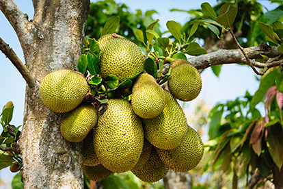
Researchers in Brazil have created a new biomaterial that could offer an effective way to treat periodontitis, a serious form of gum disease. The team at the Pontifical Catholic University of São Paulo developed the material as part of an effort to improve treatment options for a condition that can destroy the structures holding teeth in place, leading to bone loss and reduced attachment between teeth and surrounding tissues.
Current treatments for periodontitis are designed to control infection and inflammation, but they generally do little to regenerate damaged periodontal tissue. Other approaches have been explored, but their results can vary and are often difficult to predict. To overcome these limitations, the researchers investigated natural bioactive materials that could address several aspects of the disease simultaneously.
The researchers combined jackfruit latex with pomegranate peel extract, which is known for its antimicrobial properties when applied topically, and simvastatin, an anti-inflammatory drug that has been studied for its ability to stimulate bone formation. Together, the ingredients formed a mucoadhesive matrix designed to act directly on damaged tissue. The biomaterial was found to promote osteoinduction, the process that encourages cells to develop into bone-forming tissue, within 14 days. The effect became stronger after 21 days, supporting the material’s potential as a treatment for periodontitis.
Prof. Eliana Aparecida de Rezende Duek, who co-ordinated the study, said: “We observed that the developed biomaterial has great potential for future applications in treating periodontitis and in other areas as well, especially since it involves a material that has received little attention in the scientific literature for biomedical use”.
The study was published in Polymer Bulletin.
From: https://www.sciencedaily.com/releases/2026/06/260618041508.htm.
Study links periodontal disease to autoimmune disease and all-cause mortality
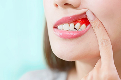
Periodontal disease may be associated with wider systemic inflammatory and immune-related health risks, according to a new population-based study by researchers at King’s College London. The study examined the relationship between periodontal disease and autoimmune disease, as well as the combined association of both conditions with all-cause mortality during follow-up. The findings add to existing evidence linking periodontal health with systemic disease and suggest that periodontal disease may serve as a clinically relevant indicator of broader health vulnerability, particularly when it occurs alongside autoimmune disease.
The study included 11,739 participants from the US and 433,023 participants from the UK. The researchers found that autoimmune diseases were more common among individuals with periodontal disease, particularly among edentulous people. Autoimmune disease was associated with higher all-cause mortality, and this risk further increased when periodontal disease was also present.
Lead author Dr Ke Zhou said: “This does not mean that periodontal disease directly causes autoimmune disease or death, but it does suggest that oral health may provide useful insight into a person’s general health status”. Dr Zhou said that the findings should be viewed in the context of established research on periodontal inflammation and systemic health and noted that periodontal disease may be connected to broader patterns of immune imbalance.
For dentistry, the findings reinforce the value of periodontal assessment as part of a broader understanding of patient health. The findings may be particularly relevant for patients with autoimmune disease, who may already have a higher inflammatory burden.
The study was published online in the Periodontology Journal.
Titanium particles linked to antibiotic failure in peri-implantitis
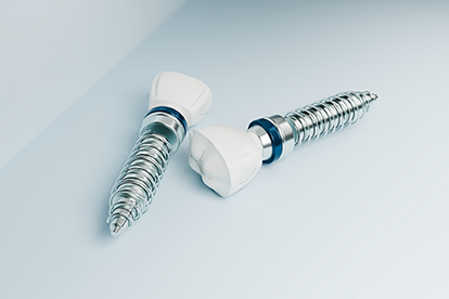
In peri-implantitis cases that persist despite standard antimicrobial therapy, it is not clear what prevents the host response from resolving the disease. Implant-derived titanium particles have been implicated in peri-implant inflammation, but how they might contribute to persistent peri-implantitis has remained unclear. A new study from Rutgers School of Dental Medicine has investigated this gap, and reports a mechanism that may help explain why antimicrobial therapy alone can be insufficient in some cases.
The study shifts attention from the microbial component of peri-implantitis to the implant surface. Bacterial biofilms can corrode titanium implant surfaces, releasing microscopic particles into the peri-implant tissue. Titanium particles can be released during maintenance if instruments intended for natural teeth are used on implants and during procedures used to treat established peri-implant inflammation. The researchers examined what these particles do after they enter peri-implant tissue. They found that the particles can interfere with macrophage function, reducing the ability of these immune cells to engulf and destroy bacteria, and promoting a sustained inflammatory response associated with bone destruction.
Senior author Prof. Georgios Kotsakis said: “For the first time, we show why all the antibiotic treatments that work around teeth do not work around implants. Now that we know the cause, we can start developing therapeutics”.
For clinicians, the findings have direct significance for implant maintenance and peri-implantitis treatment. Instrumentation that damages implant surfaces may add to the biological process that treatment is intended to control. The study therefore adds mechanistic support for implant-specific maintenance protocols and careful instrumentation.
The study was published online in PNAS Nexus.
Plant-based chewing gum reduces oral microbes associated with head and neck cancer

Oral and oropharyngeal cancers remain a significant clinical challenge, and growing evidence has linked the oral microbiome to head and neck squamous cell carcinoma (HNSCC) development, progression or recurrence. Researchers at the University of Pennsylvania School of Dental Medicine have investigated whether a plant-based chewing gum formulation can reduce HNSCC-associated oral pathogens. The findings suggest that this approach could eventually be explored as a locally delivered adjunct for lowering microbial risk factors associated with oral and oropharyngeal cancer.
The research team collected saliva and oral rinse samples from patients with HNSCC and exposed these in the laboratory to extracts prepared from the chewing gum. The chewing gum formulation contained a protein derived from lablab beans that can bind to viruses and trap them, helping to prevent their entry into host cells.
The researchers then optimised the chewing gum formulation by adding an antimicrobial peptide, seeking to target Porphyromonas gingivalis and Fusobacterium nucleatum, oral bacteria that have been implicated in oral cancer and HNSCC progression and survival outcomes. In ex vivo experiments, they found that the formulation reduced the concentration of both bacteria in saliva and oral rinse samples by more than 99%. The study also reported only minimal effects on selected non-pathogenic oral bacteria.
According to lead researcher Prof. Henry Daniell, the next step is clinical evaluation after regulatory authorisation: “Based on the successful reduction of these pathogens in ex vivo clinical studies, we plan to advance antiviral and antibacterial chewing gums for evaluation in HNSCC patients after authorisation by the US Food and Drug Administration of an investigational new drug application”.
The study was published online in Scientific Reports.


